
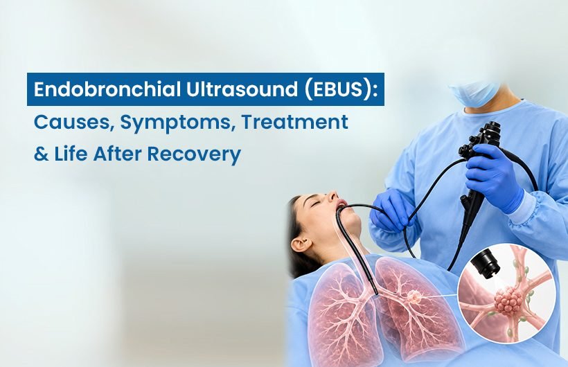
If your pulmonologist has recommended an EBUS after finding something unusual on your chest X-ray or CT scan, it’s natural to feel a little anxious about what comes next. The good news is that EBUS is one of the safest and least invasive ways to get a definite answer about what’s going on inside your chest, often without any cuts or stitches at all.
As a pulmonologist in Ahmedabad with a focus on advanced bronchoscopy and interventional lung procedures, Dr. Rushi Desai regularly uses EBUS to diagnose lung infections, inflammatory conditions, and cancer, and to work out how far a cancer has spread before deciding on treatment. This guide walks you through what EBUS actually is, the different types performed, how it differs from a standard bronchoscopy, and what risks, cost, and recovery look like.
What Is Endobronchial Ultrasound (EBUS)?
Endobronchial Ultrasound, or EBUS, is a specialised form of bronchoscopy that combines a thin, flexible camera with an ultrasound probe on its tip. The pulmonologist passes this instrument through your nose or mouth, down your windpipe, and into your airways, using the ultrasound to look through the airway wall at the lymph nodes, blood vessels, and tissue masses that sit just outside it areas a plain bronchoscope alone can never see.
Because the probe can be steered to a precise spot under live ultrasound guidance, the doctor can often take a tissue sample from that exact location in the same sitting, rather than needing a separate biopsy procedure. This is what makes EBUS so valuable for diagnosing conditions such as:
- Lung cancer and cancers that have spread to the chest lymph nodes
- Enlarged mediastinal lymph nodes of unclear cause
- Tuberculosis and other lung infections
- Sarcoidosis and similar interstitial lung disease conditions
- Unexplained lung nodules or masses seen on imaging
“EBUS has changed how we approach chest lymph nodes and hard-to-reach lung lesions,” says Dr Rushi Desai. “In many cases, it gives us a tissue diagnosis and cancer stage in a single outpatient procedure that used to require surgery under general anaesthesia.”
Types of Endobronchial Ultrasound
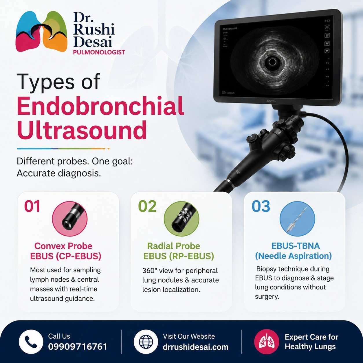
Not all EBUS procedures are the same. Depending on where the abnormality is located, your pulmonologist will choose one of the following approaches.
1. Convex Probe EBUS (CP-EBUS)
This is the most used form of EBUS. The convex probe sits at the tip of the bronchoscope and looks forward and to the side, giving a clear view of the mediastinum, the space between the lungs that houses the major lymph node stations. Its biggest advantage is that it allows EBUS-guided transbronchial needle aspiration (EBUS-TBNA): the doctor can pass a fine needle through the scope and take a biopsy from a lymph node or mass in real time, guided directly by the ultrasound image. This is the technique most often used for staging lung cancer.
2. Radial Probe EBUS (RP-EBUS)
The radial probe gives a 360-degree, high-resolution view of the airway and the tissue surrounding it. It’s especially useful for reaching smaller nodules further out in the lung, beyond where a convex probe can go. The trade-off is that radial EBUS is used mainly for locating a lesion accurately if a biopsy is needed, the doctor typically switches to a separate biopsy instrument passed through the same airway route.
3. EBUS-TBNA (Transbronchial Needle Aspiration)
Strictly speaking, this isn’t a separate “type” of scope but rather the biopsy technique performed during a convex probe EBUS. It’s specifically what allows sampling of lymph nodes and central lung masses without any incision, and it’s the technique most often used for lung cancer staging and for diagnosing conditions such as sarcoidosis and TB lymphadenitis.
What’s the Difference Between Bronchoscopy and EBUS?
Bronchoscopy vs EBUS
Both procedures use a bronchoscope, but they provide different views and are useful for different diagnostic purposes.
Standard Bronchoscopy
Direct airway examinationPrimarily provides a direct view of the inside of the airways.
Uses direct visual guidance while examining and sampling visible airway abnormalities.
Airway lesions, infections, mucus or foreign-body clearance and direct airway assessment.
EBUS
Ultrasound-guided examinationHelps visualize the airway wall and structures beyond it, including lymph nodes and nearby masses.
Uses real-time ultrasound guidance to help target tissue or lymph-node sampling.
Mediastinal lymph nodes, cancer staging and lesions located deeper around the airways.
The choice between bronchoscopy and EBUS depends on what needs to be examined or sampled. Your pulmonologist can determine which procedure is appropriate based on your symptoms, imaging findings and clinical requirements.
Why Would You Need an EBUS?
Your doctor may recommend EBUS if a chest CT or PET scan shows enlarged lymph nodes or a mass near the airways, or if you have symptoms such as a persistent cough, unexplained weight loss, or coughing up blood that need further evaluation. It’s especially central to two situations:
- Diagnosing the cause of an abnormality infection, inflammation (like sarcoidosis or other interstitial lung disease), or cancer from a single tissue sample.
- Staging lung cancer determining whether cancer has spread to lymph nodes in the chest, which directly affects whether surgery, chemotherapy, radiation, or a combination is the right treatment path.
- combination is the right treatment path.
How to Prepare for an EBUS Bronchoscopy
Preparation is similar to a standard bronchoscopy, and your care team will give you specific instructions, but in general you should expect to:
- Fast before the procedure; typically no food or water for 6–8 hours beforehand, so your stomach is empty under sedation.
- Review your medications: blood thinners, certain diabetes medications, and some other drugs may need to be paused beforehand; always tell your doctor about everything you’re taking, including supplements.
- Arrange transport home; you’ll be sedated, so you cannot drive yourself afterwards. Plan for someone to accompany you.
- Get pre-procedure tests done; bloodwork and sometimes an ECG may be requested, particularly if you have underlying heart or lung conditions.
- Ask questions upfront to clarify what sedation level to expect (moderate sedation or general anaesthesia) and how long you’ll need to stay for observation afterwards.
What Happens During and After the Procedure
EBUS is done as an outpatient procedure and typically takes 30 to 90 minutes, depending on how many lymph node stations or lesions need to be sampled. You’ll be sedated or under general anaesthesia, so you won’t feel the scope or the biopsy needle. Your throat will also be numbed with a local spray.
Afterwards, you’ll rest in a recovery area for one to two hours until the sedation wears off. It’s common to have a mild sore throat, a hoarse voice, or a slight cough for a day or so; this settles on its own. A small amount of blood-tinged mucus when coughing is also normal in the first few hours. Most people resume normal activities the following day, though your doctor will advise you on food, drink, and driving restrictions for the rest of the procedure day.
Biopsy samples are sent to the lab for analysis, and your pulmonologist will discuss the results with you at a follow-up visit, along with next steps if further treatment is needed.
results with you at a follow-up visit, along with next steps if further treatment is needed.
Risks of Endobronchial Ultrasound
EBUS is considered a very safe, minimally invasive procedure, and serious complications are uncommon. Still, as with any procedure involving sedation and airway instrumentation, it’s worth knowing the possible risks:
- Sore throat, hoarseness, or cough are the most common after-effects, usually mild and short-lived
- Minor bleeding at the biopsy site, particularly if a needle sample was taken
- Infection rare, since sterile technique is used throughout
- Pneumothorax (partial lung collapse) uncommon and more likely with certain lesion locations; may need a chest tube, or rarely a thoracoscopy, if it occurs
- Breathing difficulty or low oxygen during sedation is closely monitored and managed by the anaesthesia team throughout the procedure
- Reaction to sedation medication: your team will review your history beforehand to reduce this risk
“For most patients, the benefit of getting an accurate diagnosis, often avoiding a more invasive surgical biopsy altogether, clearly outweighs these risks,” notes Dr Rushi Desai. “We screen every patient’s history carefully beforehand so the procedure is as safe as possible.”
Why Choose Dr Rushi Desai for EBUS in Ahmedabad
Dr Rushi Desai is a bronchoscopy specialist in Ahmedabad with focused training in advanced pulmonary procedures, including EBUS, EBUS-TBNA, rigid and flexible bronchoscopy, and lung cry biopsy. As a lung transplant specialist in Ahmedabad as well, he brings a broader perspective to complex chest conditions from first diagnosis through to staging and long-term management with access to modern diagnostic technology and a patient-first approach to explaining every step of the process.
Endobronchial Ultrasound (EBUS) Care in Ahmedabad
If your doctor has recommended an EBUS, or if you’d like a second opinion on a chest CT or PET finding, book a consultation with Dr. Rushi Desai’s pulmonology clinic in Ahmedabad.
Frequently Asked Questions
Who does the EBUS biopsy?
An EBUS biopsy is performed by a pulmonologist trained in interventional bronchoscopy, typically working alongside an anaesthesia team who manages your sedation and a cytopathology team who examines the tissue samples during or after the procedure.
How much time does an EBUS test take?
Most EBUS procedures take between 30 and 90 minutes, depending on how many lymph node stations or areas need to be examined and sampled. You should also plan for one to two additional hours of recovery time before you’re discharged.
What should I eat before the EBUS procedure?
You’ll generally need to fast no food or water for about 6 to 8 hours before the procedure, since you’ll be sedated. Your doctor will confirm the exact fasting window and let you know if any regular medications should be taken with a small sip of water.
How should I prepare for an EBUS bronchoscopy?
Follow your doctor’s fasting instructions, disclose all medications and supplements you take (especially blood thinners), arrange for someone to drive you home afterwards, and complete any pre-procedure bloodwork your doctor requests. Avoid alcohol the night before if advised.
What is the success rate of EBUS biopsy?
EBUS-TBNA has a high diagnostic yield for sampling mediastinal lymph nodes and central lung masses, and it’s now considered a first-line tool for lung cancer staging in most cases. Success can vary depending on the size, location, and nature of the lesion, so your pulmonologist can give you a more specific picture based on your scans.
Is EBUS the same as a PET scan?
No. A PET scan is an imaging test that highlights areas of high metabolic activity, which can suggest cancer or inflammation, but it cannot confirm a diagnosis on its own. EBUS is a procedure that reaches the suspicious area directly and takes a tissue sample, which is what actually confirms what the abnormality is.
What is the recovery time for EBUS?
Most people go home the same day, within a couple of hours of the procedure ending, and return to normal daily activities the next day. A mild sore throat or hoarse voice for 24–48 hours is common and not a cause for concern
This article is for general information and does not replace personalised medical advice. Please consult Dr Rushi Desai or a qualified pulmonologist to discuss your specific symptoms, scan results, or need for an EBUS procedure.